index

Understand the metabolic markers that show nutrient deficiencies, mitochondrial dysfunction, infections, and toxin exposure: the test results doctors often overlook.

By Brian Wentzel | GoneGreenStore.com | Updated April 2026

What Organic Acids Are: Your Metabolic Signature

Every moment, your cells are processing nutrients, creating energy, fighting infections, and eliminating waste. Each of these biochemical processes produces byproducts (organic acids) that get eliminated through your urine.

The Organic Acids Test (OAT) captures a snapshot of these byproducts. By analyzing which organic acids are elevated or deficient in your urine, a skilled practitioner can read your metabolic story with remarkable clarity.

Think of it this way: if your bloodwork is a single photograph, your OAT is a detailed documentary of what's actually happening in your cells day-to-day.

Why Organic Acids End Up in Urine

Your body runs dozens of metabolic pathways simultaneously. Each pathway produces end-stage byproducts (organic acids) that have no further use. The body efficiently excretes these through the kidneys into the urine. Most people never measure them. But they're valuable data.

An elevated organic acid means:

  • That metabolic pathway is overactive or dysregulated
  • The nutrient feeding that pathway is either abundant or being rapidly consumed
  • A downstream problem is forcing that pathway to work overtime
  • An infection or dysbiotic organism is producing that organic acid

A low or absent organic acid might mean:

  • The metabolic pathway is impaired
  • A nutrient deficiency is blocking that pathway
  • Mitochondrial dysfunction is reducing overall metabolic capacity

Together, these patterns tell a comprehensive story.

What the OAT Test Reveals: Eight Key Windows Into Health

The OAT typically measures 60-80 different organic acids, organized into functional groups. Here are the main windows:

1. Energy Production & Mitochondrial Function

Your mitochondria are your cells' power plants. They convert nutrients (fats, carbohydrates, amino acids) into ATP, the universal energy currency. When mitochondria dysfunction, your cells can't generate sufficient energy.

Key markers:

  • Citric acid cycle intermediates (citrate, α-ketoglutarate, succinate, fumarate, malate): Elevated levels suggest the citric acid cycle (Krebs cycle) is sluggish, meaning mitochondria aren't efficiently converting nutrients to energy.
  • Pyruvate and lactate ratio: Elevated lactate with normal pyruvate suggests anaerobic metabolism, meaning your cells are working without adequate oxygen and energy, producing lactate instead of energy. This correlates with fatigue, exercise intolerance, and brain fog.
  • Malate/citrate ratio: Helps distinguish between energy production problems and other metabolic blockages.

What elevated energy markers mean:

  • Chronic fatigue syndrome (very common finding)
  • Mitochondrial disease (rare, but OAT helps screen)
  • B-vitamin deficiencies (B1, B2, B3, B5 are essential cofactors in energy pathways)
  • Magnesium or CoQ10 deficiency
  • Overtraining or chronic stress (overdriving mitochondrial capacity)
  • Mycotoxin exposure (mycotoxins directly damage mitochondria)

Restoration approach: CoQ10 (ubiquinol), carnitine, B-complex vitamins, magnesium, rest, and removal from the toxin exposure that's driving the problem.

2. Nutrient Deficiencies: The Vitamin & Mineral Picture

The OAT reveals nutritional status by measuring metabolic pathways that depend on specific nutrients.

B-vitamin deficiency markers:

  • Elevated methylmalonic acid (MMA): Indicates B12 deficiency. B12 is essential for methylation and myelin formation. Deficiency causes neurological symptoms: brain fog, peripheral neuropathy, memory problems. MMA is more specific for B12 than serum B12 alone.
  • Elevated homocysteine pathway metabolites: Indicates folate (B9) and B12 deficiency. These B vitamins are critical for methylation, the process that makes and repairs DNA, produces neurotransmitters, and regulates gene expression. Deficiency impairs all of these.
  • Elevated formiminoglutamic acid (FIGLU): Another B9 deficiency marker.
  • Elevated α-ketoglutarate and other Krebs cycle intermediates: Suggests B1 (thiamine), B2, B3, or B5 deficiency, as these vitamins are cofactors for energy production enzymes.

Other nutrient markers:

  • Elevated hydroxymethylglutarate (HMG): Suggests CoQ10 deficiency. CoQ10 is essential for mitochondrial energy production and antioxidant defense.
  • Elevated proline: Can indicate vitamin C deficiency (vitamin C is essential for collagen cross-linking).

What these mean clinically:

Vitamin deficiencies are rampant in people with mold illness, chronic infection, and compromised gut health. Many people are deficient not because they don't eat enough vegetables, but because:

  • Malabsorption: Mold illness damages the gut lining (leaky gut). CIRS patients often have severe dysbiosis. Both impair nutrient absorption.
  • Rapid depletion: Chronic infection, stress, and inflammation burn through B vitamins and magnesium at accelerated rates.
  • Genetic factors: Some people (MTHFR mutations) don't methylate or process B vitamins efficiently.

Restoration approach: The specific deficiencies determine which nutrients to supplement. B12 deficiency often requires injections or sublingual/patch forms (oral absorption is poor if intrinsic factor is compromised). Folate deficiency responds to methylfolate supplementation. Magnesium, potassium, and other minerals require targeted repletion.

3. Detoxification Capacity & Phase II Function

Your liver processes all toxins through three phases: Phase I (cytochrome P450 enzymes), Phase II (conjugation with glutathione, glycine, or other compounds), and Phase III (excretion). The OAT reveals how well Phase II is working.

Key markers:

  • Elevated phenols: Indicate poor Phase II conjugation with sulfate. Phenols are abundant in many foods (apples, almonds, green tea) and from environmental sources. If Phase II sulfation is impaired, phenols accumulate and can cause symptoms. (This is the "phenol sensitivity" phenomenon.)
  • Elevated hippuric acid: Shows Phase II glycine conjugation is working (positive sign). Low hippuric acid suggests impaired detoxification capacity.
  • Elevated p-hydroxyphenylacetic acid (HPHPA): Can indicate dysbiosis (certain dysbiotic bacteria produce this) or poor detoxification.
  • Glutathione conjugates: Directly measure Phase II glutathione conjugation. Reduced levels indicate depleted glutathione or impaired sulfation pathways.

What this means:

People with mold illness and CIRS have severely depleted glutathione and impaired detoxification. This isn't laziness or genetic bad luck; it's a direct consequence of being flooded with biotoxins and inflammatory compounds that consume Phase II capacity. As detoxification becomes impaired, toxins accumulate, driving more inflammation.

Restoration approach: Glutathione repletion (IV, nebulized, or liposomal), sulfur-containing foods (garlic, onions, cruciferous vegetables), N-acetylcysteine (NAC, which boosts glutathione synthesis), sauna therapy (supports excretion), binder support (cholestyramine, activated charcoal, which prevents reabsorption of detoxified toxins), and removal from ongoing toxin exposure.

4. Gut Dysbiosis Markers: Infections and Microbial Byproducts

Your gut microbiome is crucial. When dysbiotic organisms (harmful bacteria, yeasts, parasites) proliferate, they produce specific organic acids as byproducts.

Key markers:

  • Elevated D-lactic acid: Indicates overgrowth of lactobacillus or other lactic acid producers. Can cause brain fog, fatigue, and paradoxical reaction to fermented foods or probiotics.
  • Elevated proprionic acid, butyric acid, valeric acid, and isovaleric acid: These short-chain fatty acids (SCFAs) are normally produced by beneficial bacteria. However, elevated levels can indicate dysbiosis-driven overproduction or small intestinal bacterial overgrowth (SIBO). This correlates with gas, bloating, and neurological symptoms ("SIBO brain").
  • Elevated 2-hydroxybutyric acid: Another dysbiosis marker, often seen with SIBO.
  • Yeast metabolites: (D-arabinitol, tartaric acid, citric acid pattern): Candida and other yeasts produce characteristic organic acids. Elevated yeast markers correlate with oral thrush, vaginal yeast, systemic candidiasis, and often cognitive symptoms.
  • Hippuric acid (low): May indicate insufficient beneficial bacteria producing butyrate.
  • Elevated phenolic compounds (from dysbiotic bacterial metabolism): Dysbiotic organisms produce phenols differently than your own metabolism.

What this means:

Mold illness, CIRS, and chronic infection devastate the gut microbiome. Dysbiosis is nearly universal in these conditions. The dysbiotic microbiome further impairs detoxification, promotes systemic inflammation, and produces neuroactive compounds that worsen brain fog and mood symptoms. Dysbiosis also increases intestinal permeability ("leaky gut"), allowing bacterial lipopolysaccharides (LPS) to enter the bloodstream, driving more inflammation.

Restoration approach: Antimicrobial elimination (targeted antifungals, herbal antimicrobials, or antibiotics depending on pathogen), prebiotic feeding of beneficial bacteria (partially hydrolyzed guar gum, inulin), probiotic supplementation (specific strains beneficial after pathogen reduction), and dietary support for beneficial bacteria (soluble fiber, fermented foods once dysbiosis is under control).

5. Neurotransmitter Metabolism

Neurotransmitters (serotonin, dopamine, GABA, glutamate) regulate mood, sleep, pain perception, and cognition. The OAT reveals how well your body is producing and metabolizing these molecules.

Key markers:

  • Elevated vanillylmandelic acid (VMA) and homovanillic acid (HVA): Indicate dopamine overmetabolism or chronic stress driving excessive dopamine production. Correlates with anxiety, hypervigilance, and potential dopamine depletion over time.
  • Elevated 5-hydroxyindolacetic acid (5-HIAA): Indicates excessive serotonin breakdown. Can correlate with depression, low mood, or carcinoid syndrome (very rare). More commonly indicates either serotonin overproduction (from dysbiosis producing excessive serotonin) or rapid breakdown due to stress or genetic factors.
  • Elevated kynurenine pathway metabolites (kynurenic acid, quinolinic acid): Indicate increased tryptophan metabolism away from serotonin production. Seen in chronic inflammation and chronic stress. Quinolinic acid is neurotoxic at high levels.
  • Low GABA-related metabolites: Can indicate insufficient GABA production or excessive GABA consumption under stress.

What this means:

People with mold illness commonly show dysregulated neurotransmitter metabolism. Chronic inflammation drives excessive degradation of serotonin and dopamine, contributing to depression and mood dysregulation. Chronic stress and dysbiosis skew tryptophan metabolism away from serotonin toward the kynurenine pathway, further reducing serotonin availability. The result: mood symptoms that don't respond to SSRIs because the problem isn't serotonin signaling; it's serotonin production and availability.

Restoration approach: L-tryptophan or 5-HTP supplementation (precursors to serotonin), B6 and magnesium (cofactors for neurotransmitter synthesis), stress reduction, dysbiosis treatment, and anti-inflammatory dietary modifications. In some cases, supporting dopamine with L-tyrosine or L-DOPA precursors.

6. Oxidative Stress & Antioxidant Status

Your cells continuously produce reactive oxygen species (ROS) as metabolic byproducts. Antioxidant systems neutralize ROS. When ROS overwhelm antioxidant capacity, oxidative stress develops, a primary driver of chronic disease and aging.

Key markers:

  • Elevated isoprostanes (F2-isoprostanes, F4-neuroprostanes): Direct markers of lipid peroxidation (fat oxidation). Elevated levels indicate significant oxidative stress. Common in mold illness, CIRS, and chronic infection.
  • Elevated lipid peroxides: General markers of oxidative damage to cell membranes.
  • Elevated 8-hydroxy-2-deoxyguanosine (8-OHdG): Indicates DNA oxidative damage.
  • Low antioxidant markers: Reduced glutathione (GSH), reduced CoQ10, and low antioxidant enzyme cofactors (selenium, zinc, magnesium) indicate antioxidant depletion.

What this means:

Mycotoxins, chronic inflammation, and persistent immune activation generate excessive oxidative stress. The oxidative stress damages cell membranes and DNA, perpetuating inflammation. Many people with mold illness are caught in a vicious cycle: biotoxin exposure → oxidative stress → cellular damage → more inflammation → more oxidative stress.

Restoration approach: Antioxidant support (glutathione, liposomal vitamin C, alpha-lipoic acid, CoQ10, astaxanthin, curcumin), binder therapy (removes biotoxins, reduces oxidative burden), micronutrient repletion (selenium, zinc, manganese (cofactors for antioxidant enzymes)), and anti-inflammatory diet (reduces endogenous ROS production).

7. Amino Acid Metabolism & Protein Status

Amino acids are the building blocks of proteins, enzymes, and neurotransmitters. Dysregulated amino acid metabolism indicates either nutritional deficiency or metabolic dysfunction.

Key markers:

  • Elevated branched-chain amino acids (BCAAs: leucine, isoleucine, valine) with low aromatic amino acids (AAAs: phenylalanine, tyrosine, tryptophan): The Fischer ratio indicates malnutrition or catabolism. In severe cases, indicates hepatic dysfunction (the liver isn't properly processing amino acids).
  • Elevated urea cycle intermediates (ornithine, citrulline, arginine): Indicates the urea cycle (the body's nitrogen waste processing) is working overtime. Seen in high protein intake, muscle catabolism, or hepatic stress.
  • Low proline and hydroxyproline: Indicate vitamin C deficiency or collagen degradation (common in chronic illness).
  • Elevated taurine: Can indicate sulfur deficiency or dysbiosis.

What this means:

People with chronic mold illness often show severe amino acid dysregulation, indicating malnutrition despite eating adequately. This reflects malabsorption (damaged gut lining), increased protein turnover (muscle wasting from chronic illness), and excessive catabolism from persistent inflammation.

Restoration approach: High-quality protein intake (collagen, bone broth, grass-fed meat), amino acid supplementation (particularly BCAAs if muscle loss is occurring), gut healing to restore absorption, and anti-inflammatory therapy to reduce catabolism.

8. Mold Exposure & Mycotoxin Markers

While the OAT doesn't directly measure mycotoxins, specific organic acid patterns are highly suggestive of mycotoxin exposure.

Key markers:

  • Elevated D-arabinitol (yeast metabolite): Candida and Aspergillus species produce D-arabinitol. Elevated levels correlate with systemic fungal burden and often with mycotoxin exposure (people exposed to water-damaged buildings commonly develop both dysbiosis with Candida and mycotoxin uptake).
  • Elevated tartaric acid (another yeast metabolite): Similar significance to D-arabinitol.
  • Citric acid elevation (in specific pattern): When elevated alongside other dysbiosis markers, suggests mold-related dysbiosis.
  • Elevated fumaric acid: Can indicate mycotoxin exposure; some Aspergillus species produce fumaric acid.
  • Malate elevation pattern: When combined with energy cycle disruption and other dysbiosis markers, suggests mycotoxin-driven mitochondrial damage.

What this means:

People with confirmed mold exposure and CIRS commonly show multiple yeast metabolite elevations and energy metabolism disruption on OAT, providing supporting evidence of the mycotoxin burden even without direct mycotoxin testing.

Restoration approach: Antifungal therapy, binder support (cholestyramine removes biotoxins including mycotoxins from circulation), sauna and other drainage support, removal from ongoing exposure, and mitochondrial support.

OAT vs. Other Tests: Understanding the Complementary Panel

The OAT is powerful, but it's not a standalone test. Understanding how it fits with other assessments is crucial.

OAT vs. Blood Work: Different Windows

Bloodwork (conventional labs) measures:

  • Acute nutrient status (vitamin B12, folate serum levels)
  • Acute metabolic markers (glucose, electrolytes, liver and kidney function)
  • Structural proteins and enzymes currently in circulation
  • Current inflammation (CRP, ESR)

Bloodwork is essential but captures a narrow window.

OAT measures:

  • Functional nutrient status (are your cells actually using B12 and folate effectively?)
  • Metabolic capacity and efficiency
  • Byproducts of metabolic pathways
  • Chronic oxidative stress and inflammation (isoprostanes, etc.)

Practical example: Your serum B12 level might be normal, but your OAT might show elevated methylmalonic acid and homocysteine metabolites, indicating that despite normal serum B12, your cells aren't properly utilizing it (possibly due to MTHFR variants, malabsorption, or chronic inflammation impairing B12 transport). This explains why you're fatigued despite "normal" bloodwork.

OAT vs. HTMA: Mineral vs. Metabolic Status

Hair Tissue Mineral Analysis (HTMA) measures:

  • Mineral accumulation and depletion over 2-3 months
  • Heavy metal burden
  • Mineral ratios and metabolic rate
  • Calcium, magnesium, zinc, copper, cadmium, lead, etc.

OAT measures:

  • Metabolic byproducts and nutrient utilization
  • Functional status of metabolic pathways
  • Infection and dysbiosis markers
  • Detoxification and neurotransmitter metabolism

Practical pairing: A person might show normal mineral status on HTMA but elevated organic acid markers indicating mitochondrial dysfunction. This means minerals are present but the metabolic machinery isn't using them effectively. Or vice versa: HTMA shows magnesium depletion while OAT shows elevated energy intermediates despite normal organ function. Together, they paint a complete picture.

OAT vs. Stool Testing: Dysbiosis Assessment

Comprehensive Stool Analysis measures:

  • Beneficial bacteria populations (directly counts species)
  • Dysbiotic organisms (Klebsiella, Proteus, pathogenic bacteria)
  • Parasite presence
  • Inflammation markers (calprotectin, lactoferrin)
  • Digestion efficiency (fat, protein breakdown markers)
  • Candida antigen

OAT dysbiosis markers indicate dysbiosis is occurring but don't identify which specific organisms are present.

Practical pairing: OAT might show elevated D-lactic acid and tartaric acid (yeast overgrowth), but doesn't specify which Candida species or whether Aspergillus is present. Stool analysis can identify Candida species, but won't show how much yeast burden is present systemically. A skilled practitioner uses both.

OAT vs. Mycotoxin Testing: Different Perspectives

Urine Mycotoxin Testing (Realtime Labs, Great Plains Laboratory) measures:

  • Specific mycotoxins in urine (aflatoxins, ochratoxins, trichothecenes, fumonisins)
  • Direct evidence of mycotoxin exposure and uptake
  • Burden level and which mold species are involved

OAT shows the metabolic consequences of mycotoxin exposure (dysbiosis, mitochondrial damage, oxidative stress) but not the specific mycotoxin types.

Practical pairing: A person might have confirmed elevated mycotoxins but unclear whether they're currently being eliminated. OAT can show whether the body is actively processing and eliminating them (improved organic acid patterns over serial testing). Conversely, OAT might show evidence of mycotoxin damage even if urine mycotoxin testing is negative (because the mycotoxins may have been eliminated but the metabolic damage persists).

How to Read Your OAT Results: Making Sense of the Data

A typical OAT results report from functional labs like Great Plains Laboratory, Genova, or Doctors Data includes 60-80 markers, organized by category. Here's how to interpret them:

The Result Ranges

Most labs use three categories:

  • Low/Normal: The marker is within healthy range
  • High-Normal or Slightly Elevated: Mildly elevated, often worth addressing
  • Elevated or High: Clearly abnormal, requires intervention

Interpreting Elevated Markers: Always Consider Context

A single elevated marker can be a false positive or artifact. A pattern of elevated markers in the same category is clinically meaningful. For example:

Dysbiosis pattern (suggests real dysbiosis):

  • D-lactic acid: Elevated
  • Tartaric acid: Elevated
  • Arabinose: Elevated
  • Proprionic acid: Elevated
  • Yeast-related inflammation markers: Elevated

Random single elevated marker: Less clinically significant; may be dietary (ate fermented foods yesterday) or lab artifact.

The Ratio Patterns Matter More Than Individual Markers

Skilled practitioners look at:

  • Pyruvate:lactate ratio: High lactate with low pyruvate = anaerobic metabolism and mitochondrial dysfunction
  • Malate:citrate ratio: Helps localize where the Krebs cycle is blocked
  • Fischer ratio (BCAAs:AAAs): Low ratio indicates malnutrition; high ratio indicates possible catabolism

Ratios are more stable and reliable than individual markers.

Serial Testing Shows Trends

The real power of OAT is serial testing over time. A single test is a snapshot; two tests 3-6 months apart show whether your interventions are working.

Example trajectory:

  • First OAT (January): Elevated energy markers (Krebs cycle blockage), elevated D-lactic acid, elevated tartaric acid, low B12 markers, elevated isoprostanes (oxidative stress)
  • Interventions: Remove from mold exposure, start antifungal therapy, begin B12 injections, CoQ10 + carnitine + B-complex, sauna therapy, dysbiosis treatment
  • Second OAT (April): Energy markers normalizing, yeast markers decreased, B12 markers improving, oxidative stress markers trending down

This trajectory indicates the protocol is working and guides refinement of the next phase.

From OAT Results to Protocol: The Two-Axis Foundation Integration

The OAT reveals metabolic imbalances. The section below outlines how those findings map to the Two-Axis Foundation — for educational context only. OAT interpretation is nuanced; the patterns below are general frameworks. Always work with a qualified functional medicine practitioner to translate your specific results into a personalized protocol.

MEASURE Pillar (The OAT Phase)

The OAT itself is a MEASURE pillar intervention: it measures what's actually happening metabolically, providing precision data for all downstream interventions.

PROTECT Pillar: OAT Insights

OAT reveals what needs protection:

  • Mitochondria: Elevated energy markers often point a practitioner toward mitochondrial support (CoQ10, carnitine, reducing further toxin exposure) — ask your doctor what applies to your results
  • Detoxification capacity: Low Phase II markers indicate the liver needs protection and support (less toxic exposure, sauna, binder support)
  • Gut barrier: Dysbiosis markers indicate intestinal barrier damage needs protective intervention (L-glutamine, bone broth, avoid irritants)

PURIFY Pillar: OAT Insights

OAT reveals what needs purification:

  • Dysbiosis markers → Antifungal/antimicrobial support (discuss prescription vs. herbal options with your practitioner)
  • Elevated toxin burden patterns → Binder therapy, sauna, herbal drainage
  • Oxidative stress markers → Antioxidant drainage (glutathione, vitamin C)

RESTORE Pillar: OAT Insights

OAT reveals what needs restoration:

  • B-vitamin deficiency markers → Targeted B supplementation
  • Amino acid dysregulation → Protein support, amino acid supplementation
  • Neurotransmitter dysregulation → Precursor support (5-HTP, L-tyrosine, L-tryptophan are commonly explored — discuss with your practitioner before adding these)
  • Mitochondrial dysfunction → CoQ10, carnitine, respiratory chain support

Example: A patient's OAT shows:

  • Elevated pyruvate and lactate (mitochondrial dysfunction) → PROTECT with CoQ10, remove from toxin source
  • Elevated D-lactic and tartaric acid (Candida overgrowth) → PURIFY with antifungal
  • Low B12 and folate markers (deficiency) → RESTORE with B-complex
  • Elevated isoprostanes (oxidative stress) → PURIFY and PROTECT with glutathione and sauna

This becomes the four-pillar protocol.

The Cost-Benefit Analysis: Is OAT Testing Worth It?

OAT testing typically costs $300-500 through functional medicine labs (some insurance covers it if ordered by an MD, but coverage is inconsistent).

When OAT Testing Is Invaluable

OAT testing is often worth discussing with your physician if you have:

  • Unexplained chronic fatigue despite normal bloodwork
  • Brain fog that standard treatment doesn't resolve
  • Chronic dysbiosis or recurring yeast infections
  • Mood symptoms (anxiety, depression) resistant to standard treatment
  • Muscle pain or weakness of unclear origin
  • Digestive dysfunction with dysbiosis suspected
  • Confirmed mold exposure with uncertain metabolic impact
  • Multiple nutrient deficiencies that don't respond to supplementation

When OAT Testing May Not Be Necessary

You might skip it if:

  • You have clear, isolated mold allergy (IgE testing is sufficient)
  • You have acute infection with obvious cause
  • You're early in recovery and environmental remediation alone is working well

The Serial Testing Strategy

Many practitioners recommend:

  • Initial comprehensive OAT: $400-500
  • Follow-up OAT at 3-4 months: $350-450 (shows whether protocol is working)
  • Final confirmatory OAT at 6-12 months: $350-450 (documents recovery)

Total investment: $1,100-1,400 for comprehensive metabolic mapping and protocol validation.

Compared to:

  • Years of guessing and failed supplements: priceless
  • Unnecessary hospitalizations or interventions: expensive
  • Continued metabolic dysfunction: costs far more in quality of life

The OAT is typically one of the most cost-effective tests in functional medicine because it provides so much actionable data.

OAT Testing at Home: Practicality and Collection

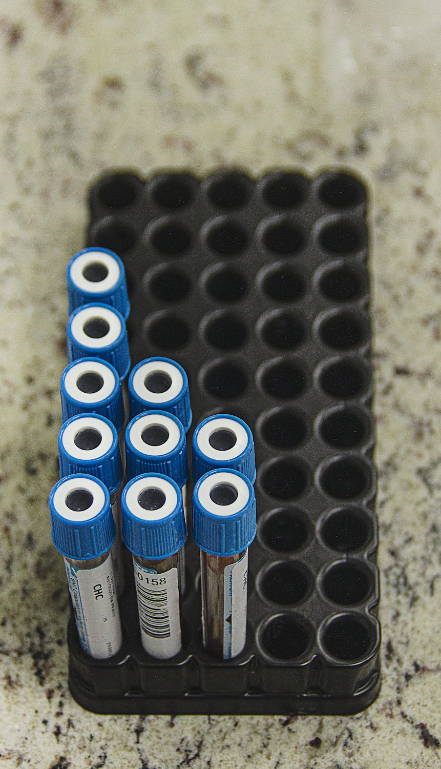
OAT testing is simple and done entirely at home:

1. Order the EquiLife OAT test kit — direct-to-consumer, no practitioner required (use code WELCOME10 for 10% off)

2. Follow collection instructions: Typically a 24-hour urine collection (sometimes first morning urine only, depends on the lab)

3. Ship the sample to the lab (prepaid shipping included)

4. Wait 2-3 weeks for results

5. Work with a practitioner to interpret results and create a protocol

No blood draws, no office visits, no special preparation. The main barrier is discipline to collect the full 24-hour sample, but most people manage it easily.

Which OAT Lab Should You Use?

Great Plains Laboratory: Comprehensive, detailed interpretation, widely recognized. $400+

Genova Diagnostics: Excellent quality, good reporting. $350+

EquiLife: Functional medicine-focused, good for mold illness and CIRS cases. $300-400

Doctors Data: Very comprehensive, medical-grade. $450+

All provide useful data. The differences are mostly in reporting detail and slight variation in normal ranges. Choose based on practitioner recommendation or cost.

Real-World OAT Patterns in Mold Illness

Based on thousands of OAT results in the mold illness community, certain patterns emerge consistently:

Classic CIRS/Mold Illness Pattern

  • Elevated pyruvate and lactate (mitochondrial dysfunction from biotoxin exposure)
  • Elevated D-lactic acid, tartaric acid, citric acid (Candida and dysbiosis secondary to mold exposure)
  • Elevated B-vitamin metabolites (malabsorption and rapid B-vitamin consumption from chronic inflammation)
  • Elevated isoprostanes and other oxidative stress markers (mycotoxin burden creating oxidative load)
  • Elevated vanillylmandelic acid or homovanillic acid (neurotransmitter dysregulation from neuroinflammation)
  • Low MSH-related markers (if available; showing immune dysregulation)
  • Impaired Phase II detoxification markers (overwhelmed detoxification from biotoxin burden)

Recovery Pattern (3-6 Months Post-Intervention)

  • Pyruvate and lactate normalizing (mitochondria recovering)
  • Dysbiosis markers decreasing (antifungal therapy working)
  • B-vitamin markers improving (absorption recovering, inflammation declining)
  • Oxidative stress markers trending down (toxin burden decreasing)
  • Neurotransmitter markers stabilizing (neuroinflammation improving)

This sequential normalization over time is reassuring and helps patients see that recovery is real, measurable, and happening.

Next Steps: Getting Your OAT

If you're dealing with mold illness, chronic fatigue, dysbiosis, or metabolic dysfunction that standard medicine hasn't solved, the OAT is one of the highest-yield tests available.

What to do next:

Order the EquiLife OAT test kit (use code WELCOME10 for 10% off; direct-to-consumer, no practitioner required)

Collect your 24-hour urine sample at home (simple, non-invasive)

Ship the sample to the lab (prepaid shipping included)

Get results interpreted — book a Lab Selection Call ($49, refundable) if you need help understanding your results

Map your specific findings to the Two-Axis Foundation (Protect, Measure, Purify, Restore protocol)

Create your precision protocol based on your actual metabolic profile, not guesswork

Re-test in 3-4 months to validate that your protocol is working and refine as needed

The OAT transforms guesswork into precision medicine. Your recovery deserves that precision.

Read next in this series:

You may also like